Tattoo my Science: Volume 1
Explore the past designs from the first iteration of the Tattoo my Science project launched in 2019
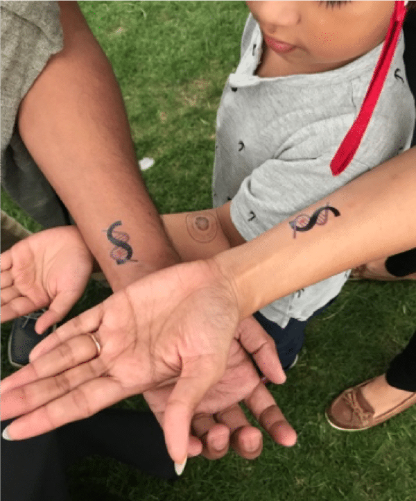
Tattoo my Science 2019:
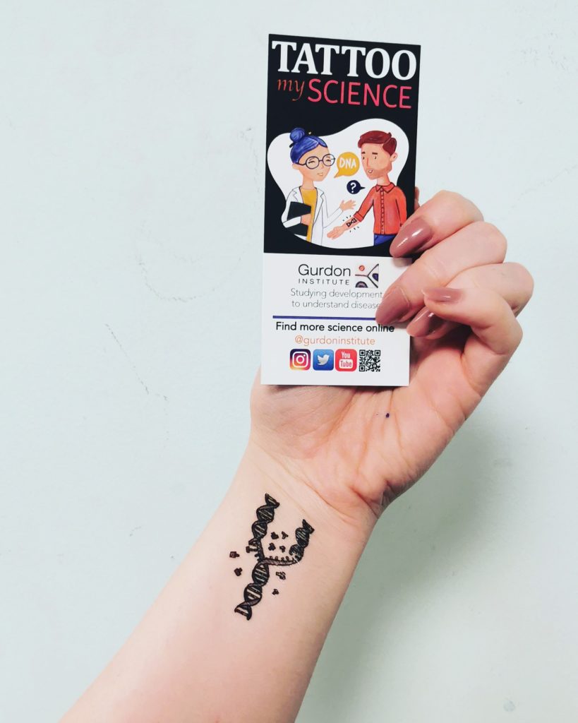
Below are some of the designs we have now retired from our collection. Either these labs are no longer at the Gurdon Institute, or they designed a brand new tattoo for the 2023 re-launch.

Circos Plot
The DNA of one cell, the egg, can direct the production of many cells, perfectly specified and organised to produce a whole animal.
Large data analysis of a cell’s DNA, the genome, can be visualised in a Circos Plot. This plot shows how different regions of the worm (C. elegans) genome interact with each other.
Design by Julie Ahringer, Ahringer lab

Fruit Fly Neurons
The human brain contains 100 billion neurons all produced by a small number of neural stem cells. As the brain grows, neural stem cells generate different cell types to produce a fully functioning nervous system.
We use the fruit fly to study how brain stem cells are organised. Remarkably, 75% of human disease causing genes are found in flies too.
Design by Jocelyn Tang, Brand Lab

DNA Repair
Every day, our cells are bombarded with agents (e.g. ultra violet light from the sun) that damage our DNA (the instructions our cells need to function). Incorrectly repaired or unrepaired DNA can lead to diseases such as cancer. Luckily, our cells have evolved multiple ways to detect and repair the damage so these mistakes are not passed onto new cells.
By studying this process, we hope to find better treatments for cancer and other diseases.
Design by Paco Muñoz-Martinez, Jackson Lab

Growing Lungs
Understanding how lung tissue naturally regenerates could offer enormous therapeutic benefits. In order to harness these benefits, we first need to understand how the human lung develops naturally.
We are studying human lung development in detail using foetal tissue and lung organoids (artificially grown masses of cells that resemble an organ in their 3D structure and function).
Design by Shuyu Liu, Rawlins Lab

Fruit Fly Egg Chamber
You may find fruit flies (aka) Drosophila on ripe fruit in your kitchen. Drosophila are used in labs at the Gurdon Institute as an animal model.
Humans and fruit flies share about 75% of the same disease-causing genes. By studying how fruit fly eggs develop, we can learn how larger animals (like whole humans) develop.
Design by Hélène Doerflinger, St Johnston Lab

Mitochondria
Each of our cells has two sets of genetic instructions: one inside the nucleus (DNA) and one inside the mitochondria (mtDNA). Mitochondria, the cell’s batteries, are tiny structures inside cells that convert the energy from food into a form that cells can use.
We study how differences in mtDNA can affect a person’s health, ageing and fertility and how these differences are inherited from mother to child.
Design by Andy Li, Ma Lab

Dementia in a Dish
Skin cells grown in a dish can be reprogrammed into stem cells and then grown into human brain cells, enabling us to study diseases in the lab, such as Alzheimer’s.
This tattoo incorporates some of the stages our cells go through in the lab, starting from skin cells on the left, the flower-like arrangement of the nerve-cell precursors (middle), and the mature neurons producing proteins involved in disease (right).
Design by Ellie Tuck, Livesey Lab

Frogs in Research
Xenopus laevis (aka the African clawed frog) is a great model animal for scientists: humans and Xenopus share many of the same disease-causing genes. Also, these frogs can produce up to 1,000 eggs in a day and their eggs are 10x bigger than human eggs (so much easier to see under the microscope). Xenopus was the first animal in the world to be cloned (by John Gurdon in 1962).
Design by Claudia Stocker, Vivid Biology

Mini Liver in a Dish
The liver and pancreas are essential organs for metabolism and energy production. We develop 3D tissue cultures, called organoids, which mimic features of these organs in a dish.
Organoids reduce the use of animals in research and also help us understand how cells regenerate. This may help us improve therapies for liver and pancreatic diseases.
Design by Olga Sarlidou, Huch Lab

Fruit Flies in Research
You may find fruit flies (aka Drosophila) on ripe fruit in your kitchen. Drosophila are used in labs at the Gurdon Institute as an animal model.
Humans and fruit flies share about 75% of the same disease-causing genes. Fruit flies are cheap to maintain. Females can lay up to 100 eggs per day and it takes only 10 days for an egg to become an adult.
Design by Claudia Stocker, Vivid Biology
Copying the Code of Life
DNA is made of four different ‘letters’, the code for building a living thing. A perfect copy of this code is made each time a cell divides, allowing organisms to grow, function, and reproduce.
We research the factors that control this copying of DNA. Some control mechanisms prevent errors which could cause cancer. Others influence how living things develop.
Design by Fiona Jenkinson, Zegerman Lab

Why Worms?
The worm known as C. elegans is a great model animal for scientists. Although worms are much simpler than humans (they don’t have bones, a heart, or a circulatory system), 35% of their genes are closely related to human genes and they share many molecular biological processes with us.
C. elegans was also the first animal to have its whole genome sequenced, in 1998.
Design by Claudia Stocker, Vivid Biology

Fluorescence
Fluorescence is an important tool in microscopy. A molecule that fluoresces (glows) has the ability to absorb light at a particular wavelength and emit light of a longer wavelength.
The first fluorescent protein used in microscopy was originally found in jelly fish.
Scientists attach fluorescent proteins to different molecules within cells in order to track their location.
Design from Thermo Fisher Scientific Fluorescence SpectraViewer bit.ly/2FaHmRh

Viral RNA
Along with DNA, RNA provides information that cells need to function. RNA comes in many different shapes and sizes. The shape of the RNA determines how it will interact with small structures inside cells (like a lock and key).
Many viruses (e.g. Zika, HIV-1 and Ebola) contain an RNA genome (instead of DNA). Understanding the shape of a virus’s RNA can help us understand how to fight the virus.
Design by Omer Ziv, Miska Lab

Cellular Potential
This is a fertilised frog embryo at the 8-cell stage.
Xenopus laevis (aka the African clawed frog) is a great model animal for scientists: humans and Xenopus share many of the same disease-causing genes. Also, these frogs can produce up to 1,000 eggs in a day and their eggs are 10x bigger than human eggs (so much easier to see under the microscope). Xenopus was the first animal in the world to be cloned (by John Gurdon in 1962).
Design by Nieuwkoop & Faber, Copyright 1994


