
Image analysis
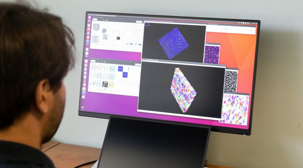
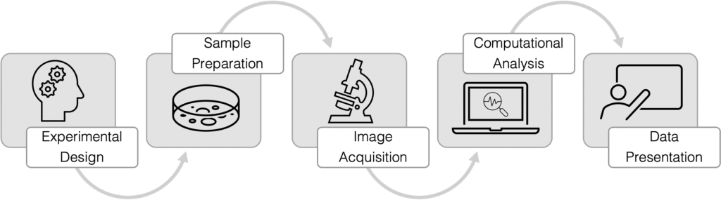
The Gurdon Institute Imaging Facility provides support with experimental design, sample preparation, image acquisition, computational image analysis and data presentation.
Our Image Analysis Specialist, Richard Butler PhD, designs pipelines and writes custom software for researchers
Previously a biologist modelling neurodegenerative disease processes, Richard is now a core programmer for the Institute specialising in developing custom image analysis software for researchers. Whether it be image data quantification, data mining, or dynamic web tools, Richard can help.
Computational image processing and analysis
Richard provides custom open-source solutions for experiments in Fiji.
Gurdon Institute Imaging Facility Fiji plugins are available to all on our GitHub page.
Alternatively, researchers have access to the commercial packages Arivis and Huygens which may be appropriate for some applications.
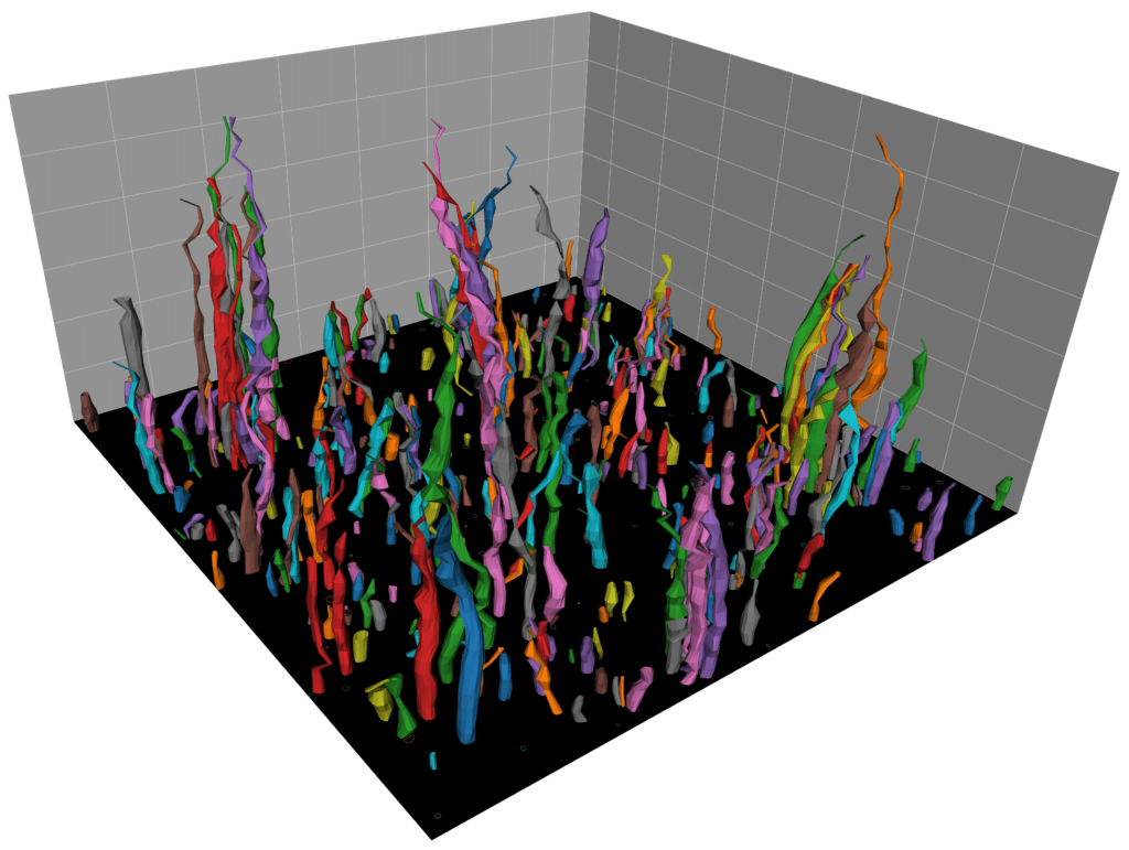
Watching filopodia grow. Three-dimensional reconstruction of filopodia-like structures growing from a supported lipid bilayer. The structures were segmented based on fluorescent actin intensity in a stack of microscopy images of size 76.13 x 76.13 x 30 microns. Colours were randomly assigned as a guide for the eye. The segmentation was performed using a custom image-analysis pipeline.