
Imaging
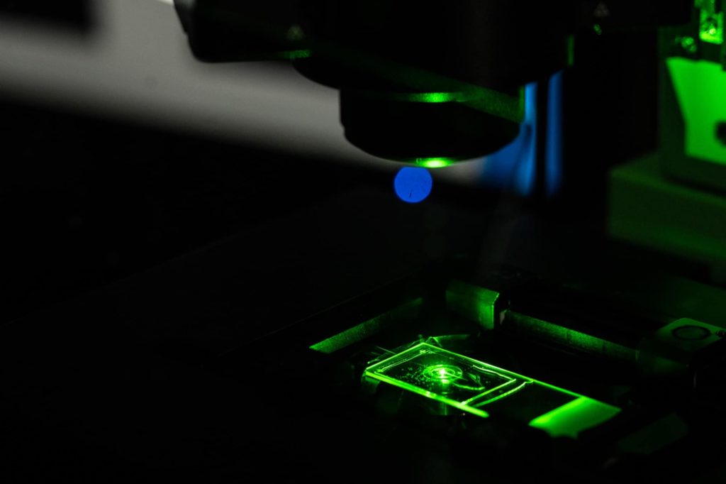
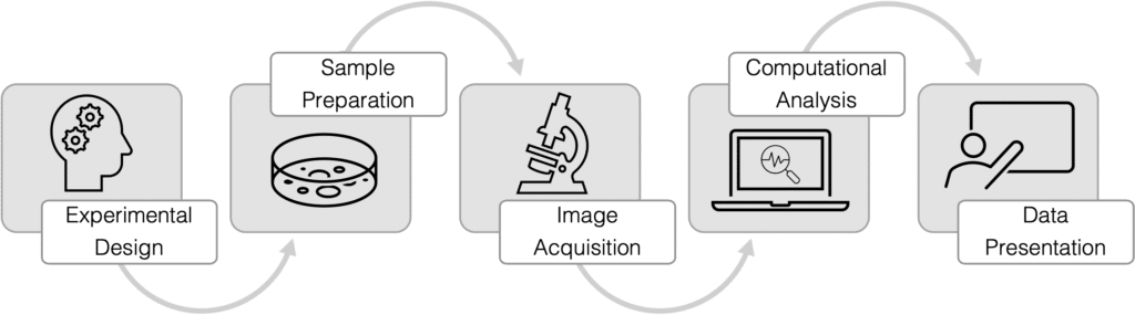
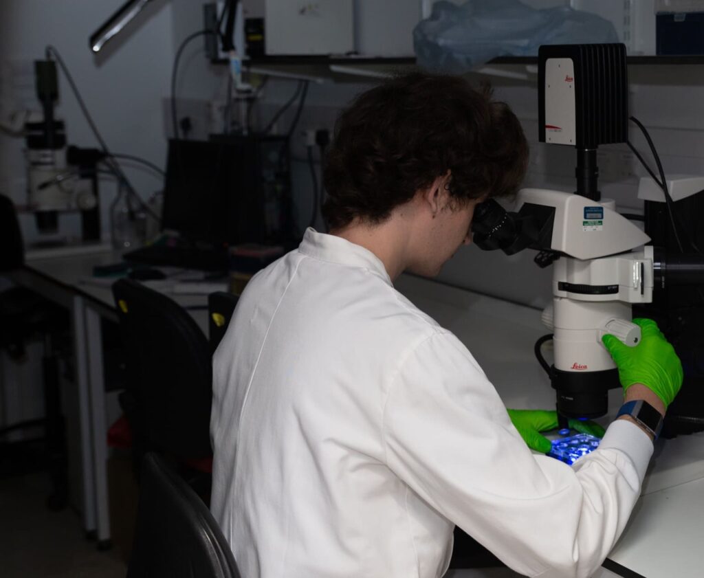
The Gurdon Institute Imaging Facility offers a wide variety of advanced light microscopy systems and provides support with experimental design, sample preparation, image acquisition, computational image analysis and data presentation as well as microscopy training
Gurdon Institute Imaging Facility microscopes
- Leica SP8 upright confocal
- Leica SP8 WL inverted confocal
- Zeiss 880 Airyscan confocal
- Zeiss Z1 Light-sheet microscope
- Nikon SoRa spinning disk
- Nikon AX R confocal
If you are interested in using one of our microscopes in your research, please head to the Gurdon Institute Imaging Facility Website for more information about arranging a visit.

Our team of imaging specialists
Nicola Lawrence PhD: Head of Imaging
With a background in developmental biology, Nicola Lawrence has extensive experience of experimental design, sample preparation and image acquisition using a wide variety of techniques from standard wide-field and confocal, to more complex methods such as structured illumination and light-sheet microscopy.
Richard Butler PhD: Image Analysis Specialist
Purnima Kumar PhD: Imaging Associate
With a research background in Virology, deciphering the roles of viral proteins by studying protein-protein interactions, Purnima has an extensive experience in various imaging techniques, from wide-field fluorescence and confocal to super-resolution microscopy.