
Zeiss Z1 light-sheet microscope
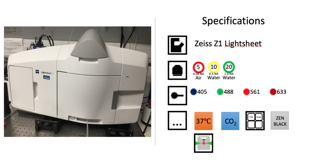
Technology focus
Light-sheet microscopy is a fluorescence technique that projects a thin ‘sheet’ of excitation light perpendicular to the direction of observation. Only the focus plane is illuminated making it gentler on the sample and thus ideal for long-term live imaging of transparent samples such as zebrafish or organoids.
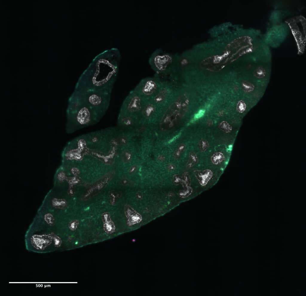
Light-sheet microscopy is also well suited to imaging larger cleared samples, such as this 6mm-cubed piece of cleared lung tissue, because of the potential imaging speed. (Sample prepared and imaged by Tessa Hughes, Rawlins lab.)