St Johnston

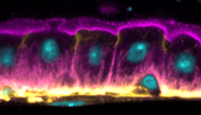
The simple layer of epithelial cells in the fruit fly midgut, with intestinal stem cells (ISC, smaller cells) situating at the basal side. Nuclear in cyan, alpha-Spectrin (cell cortical region) in magenta, and occluding septate junction in yellow.
Publication - St Johnston

How polarised epithelial cells grow “up” without breaking the barrier

Similarities between fly midgut polarisation and lumen formation in mammalian epithelial cysts

September 28, 2022

Read more
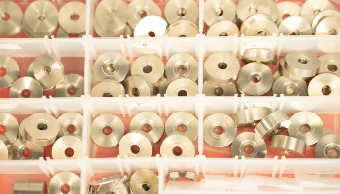
Photo of drawer full of metal microscope components
Publication - St Johnston

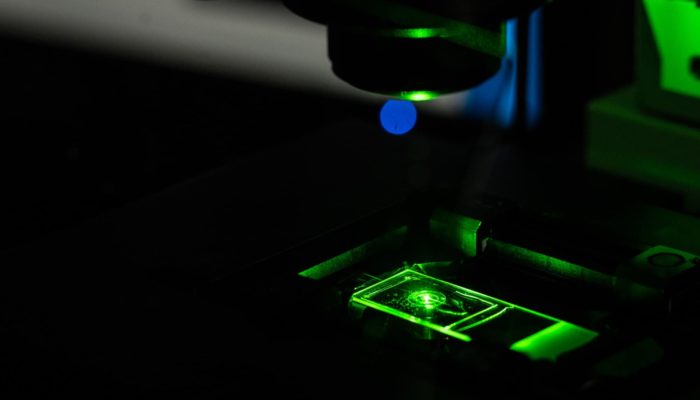
Optimising spinning disk microscopy for imaging with PAINT

Spinning disk geometries with higher light collection efficiency prove best for PAINT-based imaging

June 7, 2022

Read more
Publication - St Johnston

Unexpected clustering of nuclear pores revealed by new tissue imaging system

Single-molecule localisation microscopy in tissues reveals non-random distribution of nuclear pores

December 16, 2021

Read more
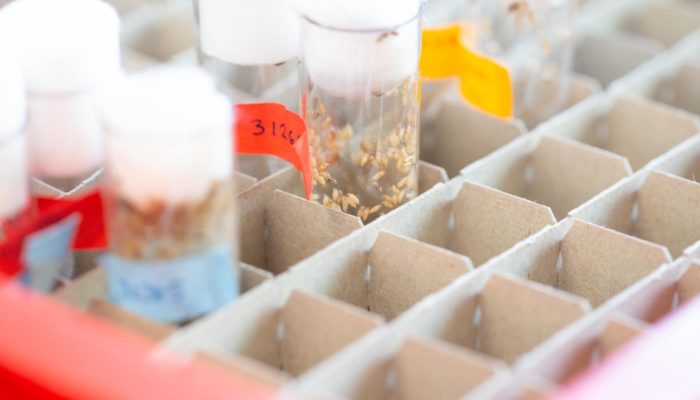
Fly vials in St Johnston lab
Publication - St Johnston

Polarity axes in Drosophila

Doerflinger et al. establish role of myosin in setting up oocyte polarity

December 1, 2021

Read more
Fly sorting in St Johnston lab
Publication - St Johnston

Breaking symmetry to establish oocyte identity

St Johnston lab show that Patronin/CAMSAP marks the future Drosophila oocyte

November 11, 2021

Read more