Human germline rejuvenation entails heterochromatin remodelling
January 18, 2023
Read more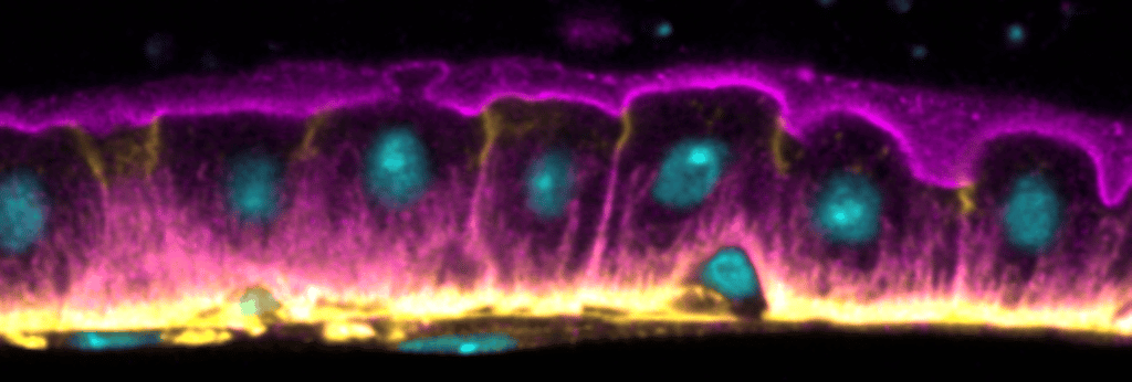
For an enteroblast to differentiate into an enterocyte, it needs grow in size, polarise and integrate between the adjacent enterocytes. Previous work has focused on the transcriptional pathways controlling enteroblast differentiation, but little was known about how the enteroblast polarises as it inserts into the epithelial layer.
This article reveals that integrating enteroblasts develop an apical domain in a similar fashion to mammalian epithelial cells in 3D culture. The process by which the cells differentiate shows other similarities with many mammalian epithelia, therefore the midgut may provide a good model for understanding how mammalian epithelia polarise. These findings may also be relevant to epithelial cancer biology, since enteroblasts that lack septate junction components fail to form an apical domain and induce the formation of tumours inside the epithelium.
Chen J & St Johnston D (2022) De novo apical domain formation inside the Drosophila adult midgut epithelium. eLife 11:e76366. DOI: 10.7554/eLife.76366.
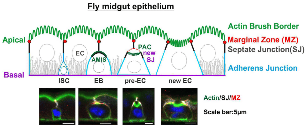
Integrating enteroblast (EB) growing "upward" (apically) and forming the Apical Membrane Initiation Site (AMIS) first, then separating the junctional (grayscale) and apical (green) components into the Pre-assembled Apical Compartment (PAC). The apical domain is formed in the newly integrated enterocyte (EC).
In the adult Drosophila midgut, basal intestinal stem cells give rise to enteroblasts that integrate into the epithelium as they differentiate into enterocytes. Integrating enteroblasts must generate a new apical domain and break through the septate junctions between neighbouring enterocytes, while maintaining barrier function.
We observe that enteroblasts form an apical membrane initiation site (AMIS) when they reach the septate junction between the enterocytes. Cadherin clears from the apical surface and an apical space appears between above the enteroblast. New septate junctions then form laterally with the enterocytes and the AMIS develops into an apical domain below the enterocyte septate junction. The enteroblast therefore forms a pre-assembled apical compartment before it has a free apical surface in contact with the gut lumen. Finally, the enterocyte septate junction disassembles and the enteroblast/pre-enterocyte reaches the gut lumen with a fully formed brush border.
The process of enteroblast integration resembles lumen formation in mammalian epithelial cysts, highlighting the similarities between the fly midgut and mammalian epithelia.