Mitoballs, large mitochondrial clusters, form to sustain sperm development
August 14, 2023
Read more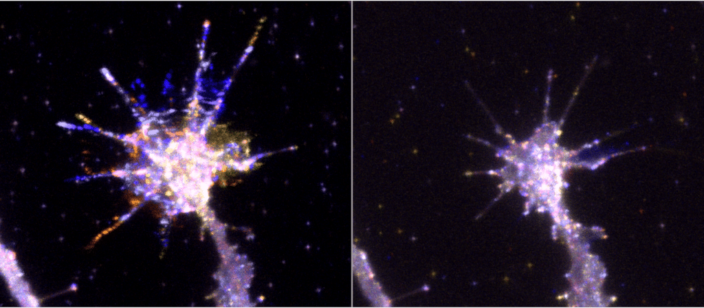
In their study, the researchers used frog retinal ganglion cells as a model to study how filopodia, dynamic cytoskeletal protrusions from the cell surface, are regulated during axon outgrowth. Using high speed imaging with quantitative dynamics analysis, they showed how particular proteins work together at filopodia. In collaboration with the Danuser lab at University of Texas Southwestern they used a novel statistical approach, Granger causality analysis. The work helps us to understand how neurons navigate accurately over long distances during brain development.
Thomas C. A. Blake, Helen M. Fox, Vasja Urbančič, Roshan Ravishankar, Adam Wolowczyk, Edward S. Allgeyer, Julia Mason, Gaudenz Danuser, and Jennifer L. Gallop (2024). Filopodial protrusion driven by density-dependent Ena-TOCA-1 interactions. J Cell Sci jcs.261057. Published online February 7, 2024. DOI: 10.1242/jcs.261057
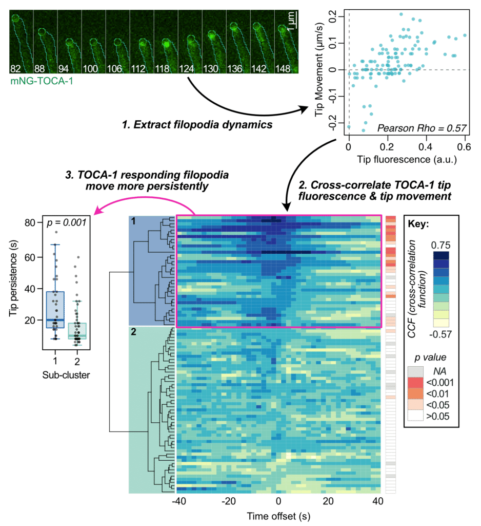
Analysis outline. TOCA-1 localises to extending filopodia tips (top left), resulting in strong correlation between tip fluorescence and tip movement (top right). Calculating cross-correlation score with different time offsets across many filopodia reveals a subset with strong correlation – ‘TOCA-1 responding filopodia’ – which have significantly higher persistence of filopodia tip movement.
Filopodia are narrow actin-rich protrusions with important roles in neuronal development where membrane-binding adaptor proteins have emerged as upstream regulators that link membrane interactions to actin regulators, for example I-BAR and F-BAR domain-containing proteins interacting with Ena/VASP and formins. To explore the significance of the F-BAR neuronal membrane adaptor TOCA-1 in filopodia we used quantitative analysis of TOCA-1 and filopodial dynamics in Xenopus retinal ganglion cells, where Ena/VASP proteins have a native role in filopodial extension. Both the adaptors and their binding partners are part of diverse and redundant protein networks that can functionally compensate for each other. Increasing density of TOCA-1 enhances Ena/VASP binding in vitro and an accumulation of TOCA-1, and its coincidence with Ena, correlates with filopodial protrusion in vivo. Two-colour single molecule localisation microscopy of TOCA-1 and Ena supports their nanoscale association. TOCA-1 clusters promote filopodial protrusion depending on a functional SH3 domain and activation of Cdc42, which we perturbed using small molecule inhibitor CASIN. We propose that TOCA-1 clusters act independently of membrane curvature to recruit and promote Ena activity for filopodial protrusion.